Litman stethoscope Amazon -50·/·OFF DEAL

https://amzn.to/3zwfAxU

cells of epidermis and their functions

 Skin Cells and Their Functions

Introduction to Skin Anatomy

- Definition and characteristics of skin

- Overview of skin layers: epidermis, dermis, hypodermis

- Importance of skin as the largest organ

 Epidermal Cells


1. Keratinocytes

   - Role in producing keratin

   - Function in forming the skin barrier

   - Differentiation process and turnover

2. Melanocytes

   - Production of melanin

   - Contribution to skin color and photoprotection

   - Regulation by UV exposure and genetics

3. Langerhans Cells

   - Function as antigen-presenting cells

   - Role in immune response within the skin

   - Interaction with other immune cells

4. Merkel Cells

   - Sensory function in touch perception

   - Location and association with nerve endings

   - Importance in tactile sensation


Dermis Cells

1. Fibroblasts

   - Synthesis of collagen and elastin fibers

   - Contribution to skin strength and elasticity

   - Role in wound healing and scar formation

2. Macrophages

   - Immune function within the dermis

   - Phagocytosis of pathogens and debris

   - Modulation of inflammatory responses

3. Adipocytes

   - Storage of energy in the form of fat

   - Thermal insulation and cushioning

   - Distribution and role in skin structure

 Hypodermis Cells

1. Adipocytes

   - Structure and function in fat storage

   - Contribution to body contour and appearance

   - Implications in metabolic health

2. Fibroblasts

   - Role in connecting dermis to underlying tissues

   - Production of collagen and elastin

   - Supportive function in skin integrity

Functions of Skin Cells

1. Protection

   - Barrier function against physical, chemical, and microbial threats

   - Role of keratinocytes, melanocytes, and immune cells

   - Importance of intact skin for overall health

2. Sensation

   - Mechanisms of touch perception and nerve signaling

   - Involvement of Merkel cells and nerve endings

   - Sensory adaptation and responses to stimuli

3. Regulation

   - Temperature regulation through sweat production

   - Role of adipocytes in thermal insulation

   - Homeostatic functions in maintaining body conditions

4. Synthesis

   - Production of structural proteins like keratin, collagen, and elastin

   - Cellular mechanisms and biochemical pathways involved

   - Implications for skin health and aging processes

Clinical Relevance and Pathological Considerations

- Common skin conditions affecting different cell types

- Diagnostic approaches and treatments

- Research advancements and future perspectives

Conclusion

- Recap of key points on skin cells and their functions

- Importance of understanding skin biology for medical practice

- Future directions in skin cell research and therapeutic interventions

Exam questions often asked from this topics are :-

1. Role of Keratinocytes in the Epidermis?

 Ans)  - Keratin production
   - Skin barrier formation
   - Keratinization process
   - Protection against damage
   - Immune function (antimicrobial peptides)

2. Significance of Melanocytes in Skin Physiology?

Ans)   - Melanin production
   - Skin color determination
   - Protection against UV radiation
   - Regulation by genetics and UV exposure

3. Langerhans Cells in the Skin's Immune System?

 Ans)  - Antigen-presenting cells
   - Detection of pathogens and allergens
   - Migration to lymph nodes
   - Initiation of immune responses

4. Functions of Fibroblasts in the Dermis?

Ans4)   - Collagen and elastin synthesis
   - Structural support
   - Wound healing
   - Contribution to scar formation

5. Role of Adipocytes in the Hypodermis?

 Ans)  - Energy storage (triglycerides)
   - Thermal insulation
   - Adipokine secretion
   - Impact on metabolic and skin health

6. Skin Cells' Contribution to Body Protection?

 Ans)  - Keratinocytes (barrier function)
   - Melanocytes (UV protection)
   - Immune cells (pathogen defense)

7. Comparison of Merkel Cells and Fibroblasts?

Ans)- Merkel cells: touch perception
   - Fibroblasts: structural support
   - Location and function differentiation

8. Implications of Dysfunctional Skin Cells in Dermatological Conditions?

 Ans)  - Examples: psoriasis, vitiligo
   - Diagnostic and therapeutic implications
   - Targeted treatments based on cell biology

9. Synthesis of Collagen and Elastin by Dermal Fibroblasts?

Ans)   - Aging effects
   - Therapeutic interventions
   - Microneedling, laser treatments, topical agents

10. Role of Skin Cells in Maintaining Homeostasis?

Ans)    - Temperature regulation (sweat, vasodilation)
    - Moisture balance (lipids, skin barrier)
    - Overall skin health support



How to loose 5kg in a month without exercise

One-Month Weight Loss Journey:

Motivation for Success

Goals:

* Define clear objectives


* Shedding pounds, fitting into clothes, improving health

Plan of Action:

*Clean up diet: whole foods, fruits, veggies, lean proteins, healthy fats

😅No eat atleast 1000 calories a day☺️


*Stay hydrated: drink plenty of water
- Increase physical activity: 30 mins most days, mix up activities


Support System:

*Surround yourself with supportive people: friends, family, online communities


Positive Mindset:

* Understand it's a journey

* Celebrate successes


*Learn from setbacks


Tracking Progress:

* Keep a food journal

* Track workouts


*Take measurements


Conclusion:

*Stay focused, positive, and motivated

*You're capable of more than you know

*Transform into the best version of yourself

ECG(EKG) interpretation

Electrocardiography (ECG)

1. Introduction to Electrocardiography:

* Non-invasive diagnostic test

*Records heart's electrical activity

* Provides insights into rhythm, rate, and conduction pathways

* Essential for diagnosing cardiac conditions


2. Basics of ECG Recording:

Electrodes Placement:
  
*10 electrodes on limbs and chest for standard 12-lead ECG

Lead Configurations:

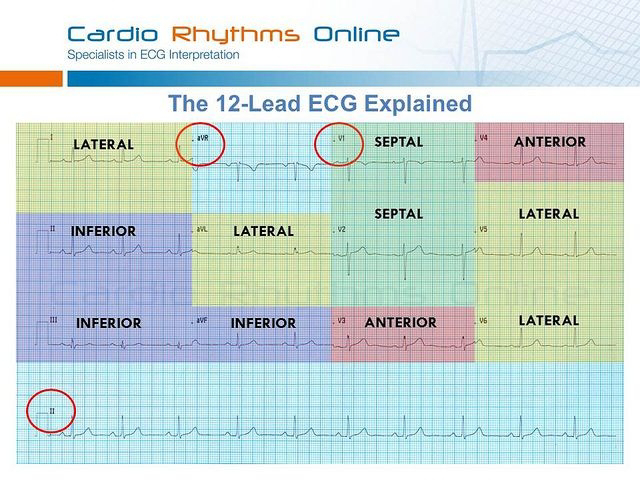
  * 12-lead system: 6 limb leads (I, II, III, aVL, aVR, aVF) and 6 precordial leads (V1-V6)

  * Each lead provides unique perspectives on cardiac function


3. Components of an ECG Waveform:
 
*P Wave: Atrial depolarization

*QRS Complex: Ventricular depolarization

 T Wave:Ventricular repolarization

*PR Interval: Atrial to ventricular depolarization time

*QT Interval:Total ventricular depolarization and repolarization time

ST Segment:Period between ventricular depolarization and repolarization, used to evaluate ischemia or injury


4. ECG Interpretation:

Normal Sinus Rhythm:Regular rhythm (60-100 bpm), P wave preceding each QRS complex, consistent PR and QT intervals

Arrhythmias: Abnormal rhythms (e.g., atrial fibrillation, ventricular tachycardia)

Myocardial Infarction(MI):
ST-segment elevation or depression, T-wave inversion indicating ischemia or infarction

ECG of MI :


Conduction Abnormalities:Prolonged PR interval, widened QRS complex, bundle branch blocks indicating conduction disturbances


5. Clinical Applications of ECG:

Diagnosis of Cardiac Conditions: Used for diagnosing myocardial infarction, arrhythmias, heart failure, electrolyte disturbances

Risk Stratification:Helps stratify patients' risk for adverse cardiac events, guides treatment decisions

Screening and Monitoring: Routine screening in patients with cardiovascular risk factors, monitoring during cardiac procedures


6. Limitations and Considerations:

False Positives and Negatives: Interpretation influenced by patient factors, technical errors, non-cardiac conditions

Supplementary Testing: Additional imaging modalities (echocardiography, stress testing, cardiac catheterization) may be required for comprehensive assessment


Conclusion:

- ECG is a fundamental tool in cardiology for evaluating cardiac function and diagnosing cardiac conditions
- Proficiency in ECG interpretation is crucial for healthcare professionals involved in cardiovascular care

Layers of skin - types , subtypes key features, diagram, histological slide view, conclusion, anatomy ,etc

Chapter Layers of Skin

Introduction:- The skin, the largest organ of the human body, plays a crucial role in protecting us from external threats, regulating body temperature, and providing sensory information. 

Comprised of multiple layers, each with distinct functions, the skin serves as a complex barrier between our bodies and the environment. 


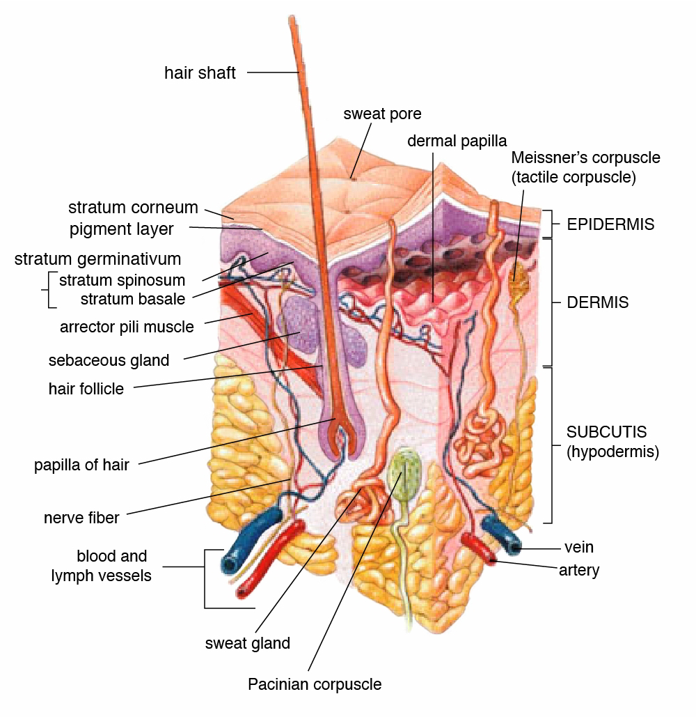
1. The Epidermis:- The epidermis is the outermost layer of the skin, primarily responsible for providing waterproofing and protection against external factors such as UV radiation, pathogens, and chemicals.

 It consists of several sublayers, including the stratum corneum, stratum lucidum, stratum granulosum, stratum spinosum, and stratum basale.


- Stratum Corneum: This is the outermost layer of the epidermis, comprised of dead skin cells called corneocytes embedded in a lipid matrix. It serves as a barrier to prevent water loss and protect against environmental damage.

Key features:- 
   - Comprised of dead skin cells called corneocytes.

   - Cells are densely packed and flattened.

   - Contains a lipid matrix that provides waterproofing and protection.

   - Acts as a barrier against water loss and external substances.

   - Regular shedding of corneocytes helps maintain skin integrity.

- Stratum Lucidum: Found only in thick skin, such as the soles of the feet and palms of the hands, this translucent layer consists of flattened keratinocytes.

Key features:-
-presents only in thick skin, such as the palms and soles.

   - Consists of translucent, flattened keratinocytes.

   - Provides additional protection and resilience to the skin.

- Stratum Granulosum: The stratum granulosum is where keratinocytes undergo terminal differentiation, forming a waterproof barrier through the release of lipids and other substances.

Key features:-
 - Contains flattened keratinocytes undergoing terminal differentiation.

   - Cells produce keratin and other proteins, contributing to the formation of the skin barrier.

   - Releases lipids and other substances that waterproof the skin.


- Stratum Spinosum: This layer contains several layers of keratinocytes connected by desmosomes, providing strength and flexibility to the skin.

Key features:-
- Consists of several layers of keratinocytes connected by desmosomes.

   - Provides strength and flexibility to the epidermis.

   - Contains Langerhans cells, which play a role in the immune response.

- Stratum Basale: Also known as the basal layer or germinative layer, this bottommost layer is responsible for the continuous renewal of the epidermis. It contains stem cells that divide and differentiate into keratinocytes, melanocytes, and Merkel cells.

Key features:-
  - Also known as the basal layer or germinative layer.

   - Contains stem cells that continuously divide and differentiate.

   - Gives rise to keratinocytes, melanocytes, and Merkel cells.

   - Melanocytes produce melanin, the pigment responsible for skin color.

   - Merkel cells function as touch receptors


2. The Dermis:- Beneath the epidermis lies the dermis, a thicker layer composed of connective tissue, blood vessels, nerves, hair follicles, and sweat glands. 

It provides structural support and elasticity to the skin and contains two main regions: the papillary dermis and the reticular dermis.

- Papillary Dermis: The superficial layer of the dermis, the papillary dermis, contains finger-like projections called dermal papillae, which interlock with the epidermis and help anchor the two layers together. It also houses sensory receptors responsible for touch and pain perception.

Key features:-
Located directly beneath the epidermis.

   - Composed of loose connective tissue.

   - Contains dermal papillae that extend into the epidermis, forming the dermal-epidermal junction.

   - Houses sensory receptors responsible for touch and pain perception.

   - Provides nutrients and oxygen to the epidermis

- Reticular Dermis: Deeper and thicker than the papillary dermis, the reticular dermis consists of dense, irregular connective tissue containing collagen and elastin fibers. These fibers provide strength and resilience to the skin, helping to prevent tearing and wrinkling.

Key features:-
 - Deeper and thicker than the papillary dermis.

   - Consists of dense, irregular connective tissue.

   - Contains collagen and elastin fibers that provide strength and elasticity to the skin.

   - Supports blood vessels, lymphatic vessels, and nerves.

   - Houses hair follicles, sebaceous glands, and sweat glands.

   - Provides structural support and resilience to the skin.


3. The Hypodermis:- Also known as the subcutaneous tissue or superficial fascia, the hypodermis is the deepest layer of the skin, consisting primarily of adipose tissue and connective tissue. 

It serves several important functions, including insulation, cushioning, and energy storage. The hypodermis also contains blood vessels and nerves that supply the skin and underlying tissues.

Key features:-
 here are the key features of the hypodermis layer:

* Adipose Tissue:
   - Predominantly composed of adipocytes (fat cells).
   - Functions as an energy reservoir, storing excess calories in the form of triglycerides.
   - Provides insulation, helping to regulate body temperature by reducing heat loss.
   - Serves as a cushion, protecting underlying organs and structures from mechanical trauma.

*Connective Tissue:
   - Consists of collagen and elastin fibers embedded in a gel-like matrix.
   - Provides support and structure to the skin and underlying tissues.
   - Connects the dermis to the underlying muscle and bone, anchoring the skin in place.
   - Allows for movement and flexibility.

* Blood Vessels and Nerves:
   - Contains blood vessels that supply oxygen and nutrients to the skin and underlying tissues.
   - Also houses lymphatic vessels that help remove waste products and toxins.
   - Contains nerves that transmit sensory information such as touch, pressure, and temperature.
   - Plays a role in thermoregulation by regulating blood flow to the skin.


Conclusion:-
The layers of the skin work together seamlessly to protect the body from external threats, maintain homeostasis, and facilitate sensory perception. Understanding the structure and function of each layer is essential for appreciating the skin's complexity and importance in overall health and well-being. By nurturing and caring for our skin, we can promote its longevity and vitality, ensuring that it continues to serve its vital functions for years to come.

Heparin Vs Warfarin Pharmacology

Heparin and warfarin  :- are both anticoagulants, but they work in different ways. Heparin works by enhancing the activity of antithrombin III, which inhibits clotting factors. 

Warfarin, on the other hand, interferes with the synthesis of clotting factors in the liver by inhibiting vitamin K epoxide reductase.


1. Mechanism of Action:-
  
 - Heparin: It works by binding to antithrombin III, which enhances its ability to inhibit clotting factors such as thrombin (factor IIa) and factor Xa. This prevents the formation of blood clots.


   - Warfarin: It inhibits the enzyme vitamin K epoxide reductase, which is necessary for the synthesis of clotting factors II, VII, IX, and X, as well as proteins C and S. By interfering with this process, warfarin reduces the production of clotting factors, thereby preventing blood clots from forming.

2. Route of Administration:-

   - Heparin: It is usually administered intravenously (IV) or subcutaneously (subQ).

   - Warfarin: It is taken orally.


3. Onset and Duration of Action:-

   - Heparin: It has a rapid onset of action but a short duration of action, typically requiring continuous infusion or frequent subQ injections.

   - Warfarin: It has a delayed onset of action and requires several days to reach therapeutic levels in the blood. Its effects are long-lasting, so it is typically taken once daily.

4. Monitoring:-

   - Heparin: Monitoring is typically done via activated partial thromboplastin time (aPTT).

   - Warfarin: Monitoring is done via international normalized ratio (INR) testing.

5. Reversal:-

   - Heparin: Its effects can be rapidly reversed with protamine sulfate.

   - Warfarin: Its effects can be reversed with vitamin K and/or specific reversal agents like phytonadione (vitamin K1) and prothrombin complex concentrate (PCC).

6. Indications:-

   - Heparin: It is often used for acute management of thromboembolic disorders, such as pulmonary embolism and deep vein thrombosis, as well as during surgeries to prevent clot formation.

   - Warfarin: It is commonly used for long-term anticoagulation in conditions such as atrial fibrillation, venous thromboembolism, and mechanical heart valve replacement.

7. Side Effects:-

   - Both drugs can increase the risk of bleeding, but heparin is associated with a higher risk of heparin-induced thrombocytopenia (HIT), while warfarin carries a risk of skin necrosis and other rare complications.


Overall, while both drugs are effective anticoagulants, their mechanisms of action, routes of administration, monitoring requirements, and indications differ, making them suitable for different clinical scenarios.

These Notes are for doctor's and medical students.

Disease of external ear detailed notes

External Ear Diseases in ENT The external ear, comprising the auricle (pinna) and the ear canal (external auditory canal), is susceptible to...