Introduction:- The skin, the largest organ of the human body, plays a crucial role in protecting us from external threats, regulating body temperature, and providing sensory information.
Comprised of multiple layers, each with distinct functions, the skin serves as a complex barrier between our bodies and the environment.

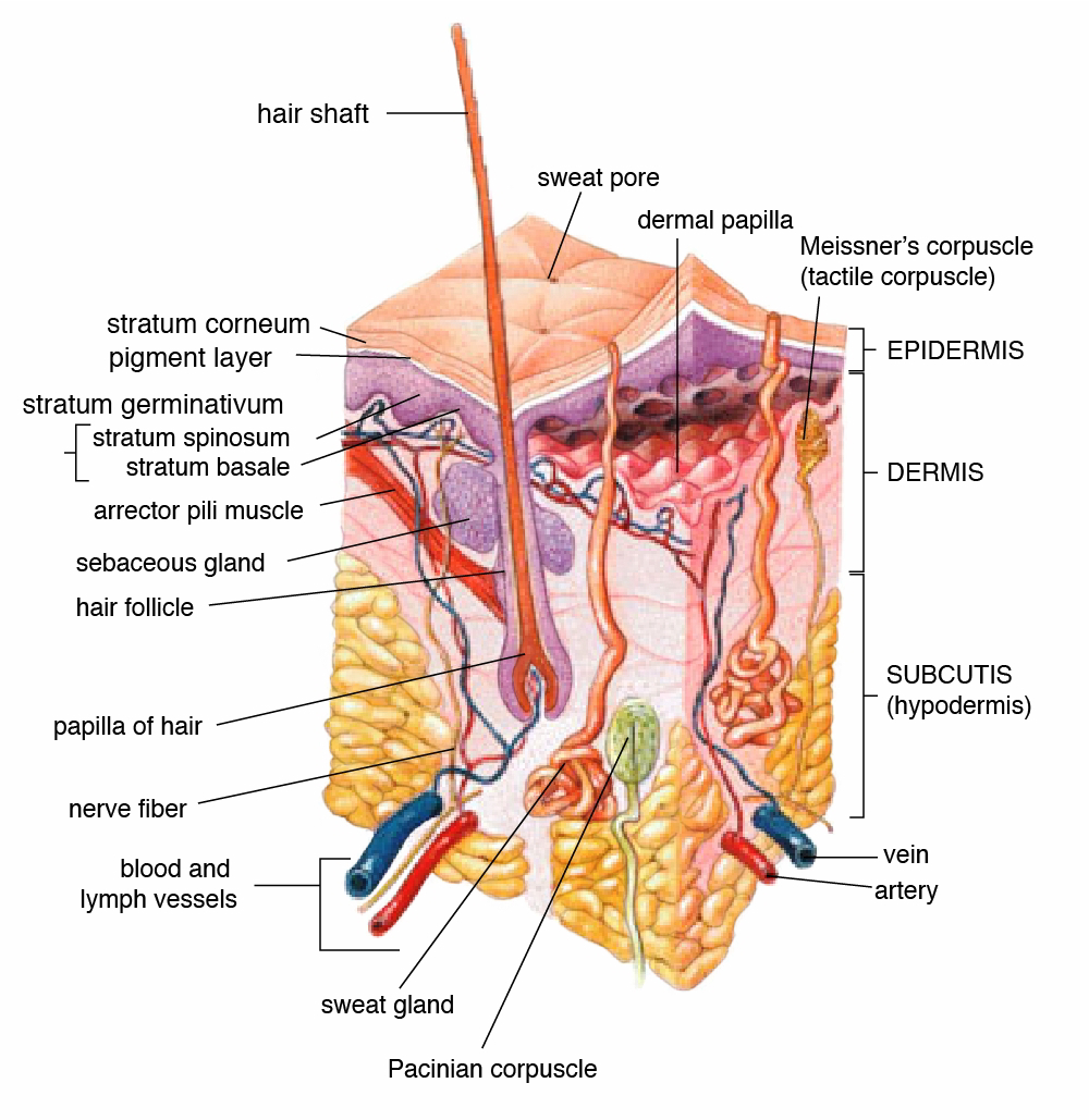
1. The Epidermis:- The epidermis is the outermost layer of the skin, primarily responsible for providing waterproofing and protection against external factors such as UV radiation, pathogens, and chemicals.
It consists of several sublayers, including the stratum corneum, stratum lucidum, stratum granulosum, stratum spinosum, and stratum basale.

- Stratum Corneum: This is the outermost layer of the epidermis, comprised of dead skin cells called corneocytes embedded in a lipid matrix. It serves as a barrier to prevent water loss and protect against environmental damage.
Key features:-
- Comprised of dead skin cells called corneocytes.
- Cells are densely packed and flattened.
- Contains a lipid matrix that provides waterproofing and protection.
- Acts as a barrier against water loss and external substances.
- Regular shedding of corneocytes helps maintain skin integrity.
- Stratum Lucidum: Found only in thick skin, such as the soles of the feet and palms of the hands, this translucent layer consists of flattened keratinocytes.
Key features:-
-presents only in thick skin, such as the palms and soles.
- Consists of translucent, flattened keratinocytes.
- Provides additional protection and resilience to the skin.
- Stratum Granulosum: The stratum granulosum is where keratinocytes undergo terminal differentiation, forming a waterproof barrier through the release of lipids and other substances.
Key features:-
- Contains flattened keratinocytes undergoing terminal differentiation.
- Cells produce keratin and other proteins, contributing to the formation of the skin barrier.
- Releases lipids and other substances that waterproof the skin.
- Stratum Spinosum: This layer contains several layers of keratinocytes connected by desmosomes, providing strength and flexibility to the skin.
Key features:-
- Consists of several layers of keratinocytes connected by desmosomes.
- Provides strength and flexibility to the epidermis.
- Contains Langerhans cells, which play a role in the immune response.
- Stratum Basale: Also known as the basal layer or germinative layer, this bottommost layer is responsible for the continuous renewal of the epidermis. It contains stem cells that divide and differentiate into keratinocytes, melanocytes, and Merkel cells.
Key features:-
- Also known as the basal layer or germinative layer.
- Contains stem cells that continuously divide and differentiate.
- Gives rise to keratinocytes, melanocytes, and Merkel cells.
- Melanocytes produce melanin, the pigment responsible for skin color.
- Merkel cells function as touch receptors

2. The Dermis:- Beneath the epidermis lies the dermis, a thicker layer composed of connective tissue, blood vessels, nerves, hair follicles, and sweat glands.
It provides structural support and elasticity to the skin and contains two main regions: the papillary dermis and the reticular dermis.
- Papillary Dermis: The superficial layer of the dermis, the papillary dermis, contains finger-like projections called dermal papillae, which interlock with the epidermis and help anchor the two layers together. It also houses sensory receptors responsible for touch and pain perception.
Key features:-
Located directly beneath the epidermis.
- Composed of loose connective tissue.
- Contains dermal papillae that extend into the epidermis, forming the dermal-epidermal junction.
- Houses sensory receptors responsible for touch and pain perception.
- Provides nutrients and oxygen to the epidermis
- Reticular Dermis: Deeper and thicker than the papillary dermis, the reticular dermis consists of dense, irregular connective tissue containing collagen and elastin fibers. These fibers provide strength and resilience to the skin, helping to prevent tearing and wrinkling.
Key features:-
- Deeper and thicker than the papillary dermis.
- Consists of dense, irregular connective tissue.
- Contains collagen and elastin fibers that provide strength and elasticity to the skin.
- Supports blood vessels, lymphatic vessels, and nerves.
- Houses hair follicles, sebaceous glands, and sweat glands.
- Provides structural support and resilience to the skin.


3. The Hypodermis:- Also known as the subcutaneous tissue or superficial fascia, the hypodermis is the deepest layer of the skin, consisting primarily of adipose tissue and connective tissue.
It serves several important functions, including insulation, cushioning, and energy storage. The hypodermis also contains blood vessels and nerves that supply the skin and underlying tissues.
Key features:-
here are the key features of the hypodermis layer:
* Adipose Tissue:
- Predominantly composed of adipocytes (fat cells).
- Functions as an energy reservoir, storing excess calories in the form of triglycerides.
- Provides insulation, helping to regulate body temperature by reducing heat loss.
- Serves as a cushion, protecting underlying organs and structures from mechanical trauma.
*Connective Tissue:
- Consists of collagen and elastin fibers embedded in a gel-like matrix.
- Provides support and structure to the skin and underlying tissues.
- Connects the dermis to the underlying muscle and bone, anchoring the skin in place.
- Allows for movement and flexibility.
* Blood Vessels and Nerves:
- Contains blood vessels that supply oxygen and nutrients to the skin and underlying tissues.
- Also houses lymphatic vessels that help remove waste products and toxins.
- Contains nerves that transmit sensory information such as touch, pressure, and temperature.
- Plays a role in thermoregulation by regulating blood flow to the skin.
Conclusion:-
The layers of the skin work together seamlessly to protect the body from external threats, maintain homeostasis, and facilitate sensory perception. Understanding the structure and function of each layer is essential for appreciating the skin's complexity and importance in overall health and well-being. By nurturing and caring for our skin, we can promote its longevity and vitality, ensuring that it continues to serve its vital functions for years to come.


