*Joints can be classified by the type of the tissue present (fibrous, cartilaginous or synovial),
*By the degree of movement permitted (synarthrosis, amphiarthrosis or diarthrosis).

Fibrous Joints
A fibrous joint is where the bones are bound by a tough, fibrous tissue. These are typically joints that require strength and stability over range of movement.
Fibrous joints can be further sub-classified into :-
*sutures
* gomphoses
*syndesmoses.
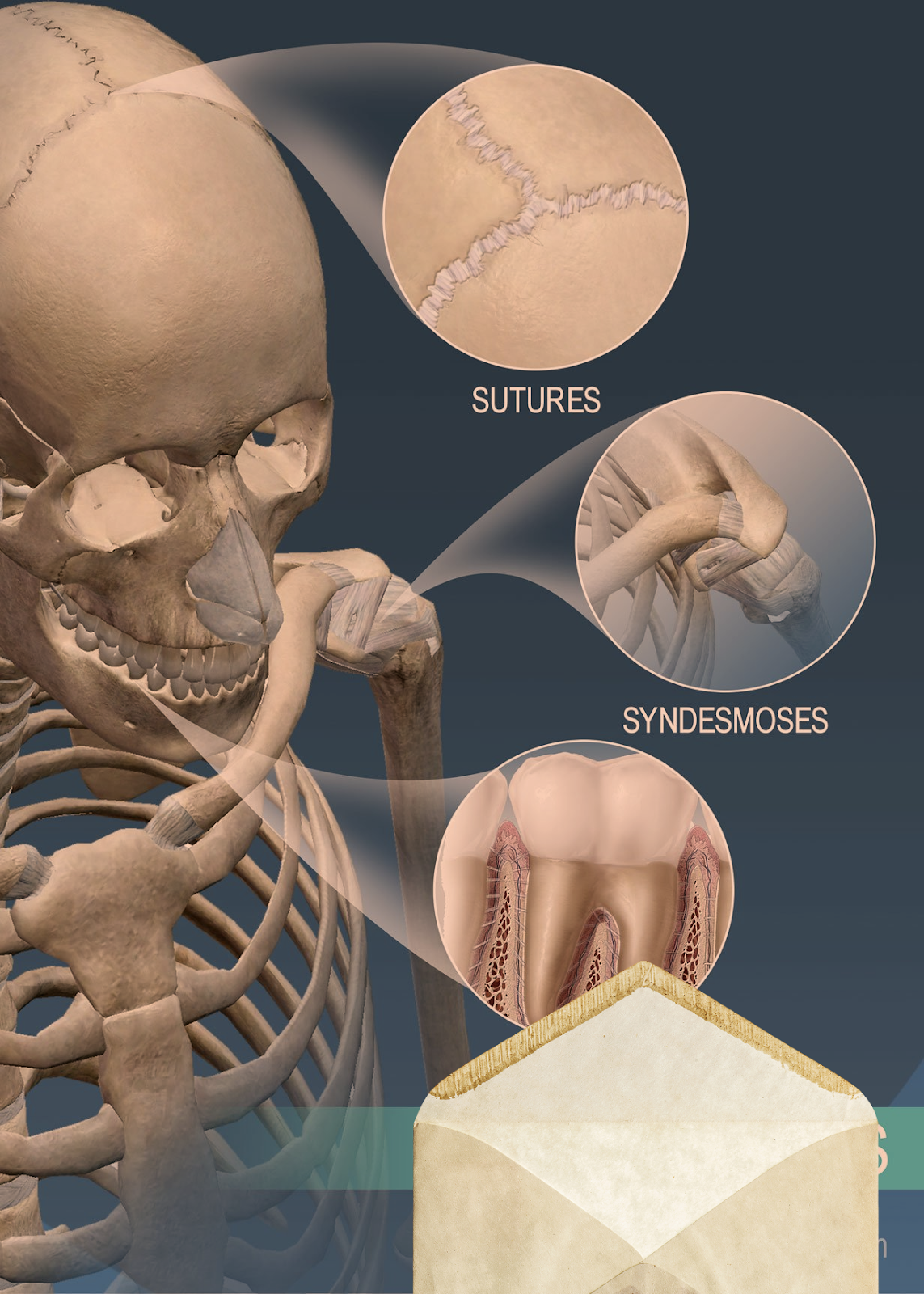
Sutures
Sutures are immovable joints (synarthrosis), and are only found between the flat, plate-like bones of the skull.
There is limited movement until about 20 years of age, after which they become fixed and immobile. They are most important in birth, as at that stage the joints are not fused, allowing deformation of the skull as it passes through the birth canal

Gomphoses
*Gomphoses are also immovable joints.
*They are found where the teeth articulate with their sockets in the maxilla (upper teeth) or the mandible (lower teeth).
*The tooth is bound into its socket by the strong periodontal ligament.
Syndesmoses
*Syndesmoses are slightly movable joints (amphiarthroses).
*They are comprised of bones held together by an interosseous membrane.
*The middle radioulnar joint and middle tibiofibular joint are examples of a syndesmosis joint.
Cartilaginous
*In a cartilaginous joint, the bones are united by fibrocartilage or hyaline cartilage.
*There are two main types: synchondroses (primary cartilaginous) and symphyses (secondary cartilaginous).
Synchondroses
*In a synchondrosis, the bones are connected by hyaline cartilage. These joints are immovable (synarthrosis).
*An example of a synchondrosis is the joint between the diaphysis and epiphysis of a growing long bone.
Symphyses
*Symphysial joints are where the bones are united by a layer of fibrocartilage. They are slightly movable (amphiarthrosis).
*Examples include the pubic symphysis, and the joints between vertebral bodies.
Synovial
*A synovial joint is defined by the presence of a fluid-filled joint cavity contained within a fibrous capsule.
*They are freely movable (diarthrosis) and are the most common type of joint found in the body.


Synovial joints can be sub-classified into several different types, depending on the shape of their articular surfaces and the movements permitted:
*Hinge – permits movement in one plane – usually flexion and extension.
E.g. elbow joint, ankle joint, knee joint.
*Saddle – named due to its resemblance to a saddle on a horse’s back. It is characterised by opposing articular surfaces with a reciprocal concave-convex shape.
E.g. carpometacarpal joints.
*Plane – the articular surfaces are relatively flat, allowing the bones to glide over one another.
E.g. acromioclavicular joint, subtalar joint.
*Pivot – allows for rotation only. It is formed by a central bony pivot, which is surrounded by a bony-ligamentous ring
E.g. proximal and distal radioulnar joints, atlantoaxial joint.
*Condyloid – contains a convex surface which articulates with a concave elliptical cavity. They are also known as ellipsoid joints.
E.g. wrist joint, metacarpophalangeal joint, metatarsophalangeal joint.
*Ball and Socket – where the ball-shaped surface of one rounded bone fits into the cup-like depression of another bone. It permits free movement in numerous axes.
E.g. hip joint, shoulder joint

Structures of a Synovial Joint
The three main features of a synovial joint are: (i) articular capsule, (ii) articular cartilage, (iii) synovial fluid.

*Articular Capsule
*The articular capsule surrounds the joint and is continuous with the periosteum of articulating bones.
*It consists of two layers:
*Fibrous layer (outer) – consists of white fibrous tissue, known the capsular ligament. It holds together the articulating bones and supports the underlying synovium.
*Synovial layer (inner) – a highly vascularised layer of serous connective tissue. It absorbs and secretes synovial fluid, and is responsible for the mediation of nutrient exchange between blood and joint. Also known as the synovium.
Articular Cartilage
*The articulating surfaces of a synovial joint (i.e. the surfaces that directly contact each other as the bones move) are covered by a thin layer of hyaline cartilage.
*The articular cartilage has two main roles: (i) minimising friction upon joint movement, and (ii) absorbing shock.
Synovial Fluid
*The synovial fluid is located within the joint cavity of a synovial joint. It has three primary functions:
*Lubrication
*Nutrient distribution
*Shock absorption
Articular cartilage is relatively avascular, and is reliant upon the passive diffusion of nutrients from the synovial fluid.
Accessory Structures of a Synovial Joint
Accessory Ligaments
*The accessory ligaments are separate ligaments or parts of the joint capsule.
*They consist of bundles of dense regular connective tissue, which is highly adapted for resisting strain. This resists any extreme movements that may damage the joint.
Bursae
*A bursa is a small sac lined by synovial membrane, and filled with synovial fluid.
*Bursae are located at key points of friction in a joint.
*They afford joints greater freedom of movement, whilst protecting the articular surfaces from friction-induced degeneration
*They can become inflamed following infection or irritation by over-use of the joint (bursitis).
Innervation
*Synovial joints have a rich supply from articular nerves.
*The innervation of a joint can be determined using Hilton’s Law – ‘the nerves supplying a joint also supply the muscles moving the joint and the skin covering their distal attachments.’

*Articular nerves transmit afferent impulses, including proprioceptive (joint position) and nociceptive (pain) sensation
Vasculature
*Arterial supply to synovial joints is via articular arteries, which arise from the vessels around the joint.
* The articular arteries are located within the joint capsule, mostly in the synovial membrane.
*A common feature of the articular arterial supply is frequent anastomoses (communications) in order to ensure a blood supply to and across the joint regardless of its position.
* In practice this usually means arteries are above and below a joint, curving round each side of it and joining via small connecting vessels.
*The articular veins accompany the articular arteries and are also found in the synovial membrane.
Osteoarthritis

*Osteoarthritis is the most common form of joint inflammation (arthritis).
*It stems from heavy use of articular joints over the course of many years, which can result in the wearing away of articular cartilage, and often the erosion of the underlying articulating surfaces of bones as well.
*The changes which occur are irreversible and degenerative.
* This results in the decreased effectiveness of articular cartilage as a shock absorber and lubricated surface, as well as the roughened edges causing further damage.
*As a result of this degeneration, repeated friction can cause symptoms of joint pain, stiffness and discomfort.
*This condition usually affects joints that support full body weight, such as the hips and the knees.
Arthritis can also come about through other causes, including; (i) as a result of infection, due to the ease with which blood (and any associated bacteria) can enter the joint cavity via the synovial membrane; (ii) due to autoinflammatory causes, as in rheumatoid arthritis, or; (iii) as a result of infection but not involving infection of the joint itself, as in reactive arthritis.


